Shedding Light on Gut Defense: A New Discovery in Crohn’s Disease

In a groundbreaking study led by postdoctoral researcher Dr. Elena Layunta , we have unraveled a critical mystery behind Crohn’s disease (CD), a debilitating inflammatory bowel condition. Our research shines a spotlight on a key molecule, MUC17, and its role in protecting the gut lining.

What Is MUC17?

MUC17 is a membrane-bound mucin, a sticky protein that forms part of the glycocalyx—a sugary barrier that coats the small intestine. This barrier acts as the body’s first line of defense, keeping harmful bacteria away from the gut lining. Think of it as the gut's natural armor.

The Breakthrough Discovery

The study revealed that MUC17 levels are significantly reduced in the small intestines of patients with Crohn’s disease. This loss weakens the glycocalyx, allowing bacteria to come into direct contact with the gut lining. Such breaches may be the initial trigger for the chronic inflammation seen in CD.

Using advanced genetic models in mice, we demonstrated that when MUC17 is absent, the small intestine becomes highly vulnerable to infections and bacterial invasion. This leads to gut barrier dysfunction and even the movement of bacteria into other parts of the body, which can contribute to further complications.

Why Does This Matter?

This discovery provides crucial insight into the early stages of Crohn’s disease, showing that disruptions in the gut’s glycocalyx barrier could occur before inflammation sets in. By identifying MUC17 as a critical player, the study opens up new possibilities for early diagnosis and targeted therapies.

Looking Ahead

This work not only advances our understanding of Crohn’s disease but also highlights the importance of the glycocalyx in maintaining gut health. Future research will explore ways to boost MUC17 levels or strengthen the glycocalyx as potential treatments for Crohn’s disease and similar conditions.

Elena’s work exemplifies how basic research can unlock new frontiers in medicine, offering hope to patients living with Crohn’s disease. Stay tuned as this exciting field of study continues to unfold!

Read the study in JCI Insight.

New paper: Intracellular transport of large transmembrane mucins

We often think of the gut as a simple passageway for food. But it’s far more than that—a highly sophisticated fortress of barriers and defenses protecting us from harmful invaders. One of its most critical components is the glycocalyx, a sugary coat lining the intestinal cells, keeping bacteria at bay.

In a fascinating new study, led by PhD student Sofia Jäverfelt, we have unraveled how a key molecule, MUC17, is transported and anchored to the gut lining. This discovery not only deepens our understanding of gut biology but also sheds light on potential causes of intestinal disorders.

What Is MUC17 and Why Does It Matter?

MUC17 is a membrane-bound mucin, a sticky protein that forms the glycocalyx layer. It acts as a shield, preventing bacteria from attaching to the gut lining and causing infections. But getting MUC17 to the right place in the gut is no simple feat—it requires a highly coordinated cellular transport system.

Sofia identified two motor proteins, MYO1B and MYO5B, as well as a sorting protein, SNX27, as the key players in ensuring MUC17 reaches and stays at the intestinal surface. If this system breaks down, the gut’s protective barrier could be compromised, leaving us vulnerable to bacterial invasion.

A Closer Look at the Findings

  • MYO1B and MYO5B: These motor proteins help transport MUC17 to the surface of gut cells. While MYO1B regulates how much MUC17 is present, MYO5B ensures it’s correctly positioned. Without these proteins, MUC17 either accumulates inside the cell or gets misplaced.

  • SNX27: This sorting protein acts like a traffic controller, directing MUC17 to its final destination and stabilizing it once it’s there. When SNX27 is missing, the mucin’s protective effects are diminished.

  • Implications for Gut Health: Defects in this transport system could explain some intestinal disorders, such as chronic infections or inflammation, by leaving the gut lining exposed to harmful bacteria.

Why This Matters

Our research provides crucial insights into how the gut maintains its defenses. Beyond its scientific impact, it offers hope for new therapies to treat or prevent conditions where this barrier fails, such as inflammatory bowel disease (IBD) or bacterial infections.

Spearheaded by Sofia Jäverfelt, this study exemplifies the importance of basic research in solving big biomedical challenges. The gut is no longer a black box—it’s a complex, finely tuned machine, and now, we’re a step closer to understanding its inner workings.

Read the paper in Biochemical Journal.

New paper on How a Tiny Protein Weakens Gut Defenses

In a groundbreaking study, our group has uncovered how a small protein on the surface of a gut-dwelling bacterium, Brachyspira pilosicoli, could hold the key to understanding some troubling intestinal conditions. This bacterium is notorious for causing intestinal spirochetosis, which is linked to symptoms like diarrhea, abdominal pain, and weight loss.

The Culprit: BPP43_05035

At the heart of the discovery is a protein called BPP43_05035, which acts as an "adhesin" – a sticky molecule that allows bacteria to latch onto cells in the gut lining. Think of it as Velcro for bacteria! But its role goes beyond simple attachment.

Using advanced tools like protein structure mapping and cell culture experiments, postdoc Anandi Rajan found that this protein doesn't just stick to the cells lining the gut; it also targets tight junctions, the cell "gatekeepers" that maintain the integrity of the intestinal barrier. By weakening these tight junctions, BPP43_05035 increases the permeability of the gut lining, allowing harmful substances and bacteria to potentially seep through.

Why Does This Matter?

The gut lining is essential for health, acting as a protective barrier against harmful microbes while letting in nutrients. When this barrier is compromised, it can lead to conditions like irritable bowel syndrome (IBS) or even systemic infections.

Our study also revealed that in patients infected with Brachyspira pilosicoli, certain protective genes in the gut lining are less active. This means the bacteria and its adhesin, BPP43_05035, could play a role in chronic gut disorders.

A Glimmer of Hope

There’s exciting potential here for medical science. Blocking the function of BPP43_05035 with specific antibodies stopped the bacteria from attaching to gut cells in lab experiments. This opens the door for targeted therapies to prevent or treat infections caused by Brachyspira pilosicoli.

What’s Next?

This discovery is a significant step toward understanding how bacteria interact with our gut. Future research will aim to develop interventions that can block bacterial adhesins like BPP43_05035 and restore gut health in affected patients.

Understanding this tiny but mighty protein could lead to big advances in managing gut health and tackling persistent infections!

Read the paper at Gut Microbes.

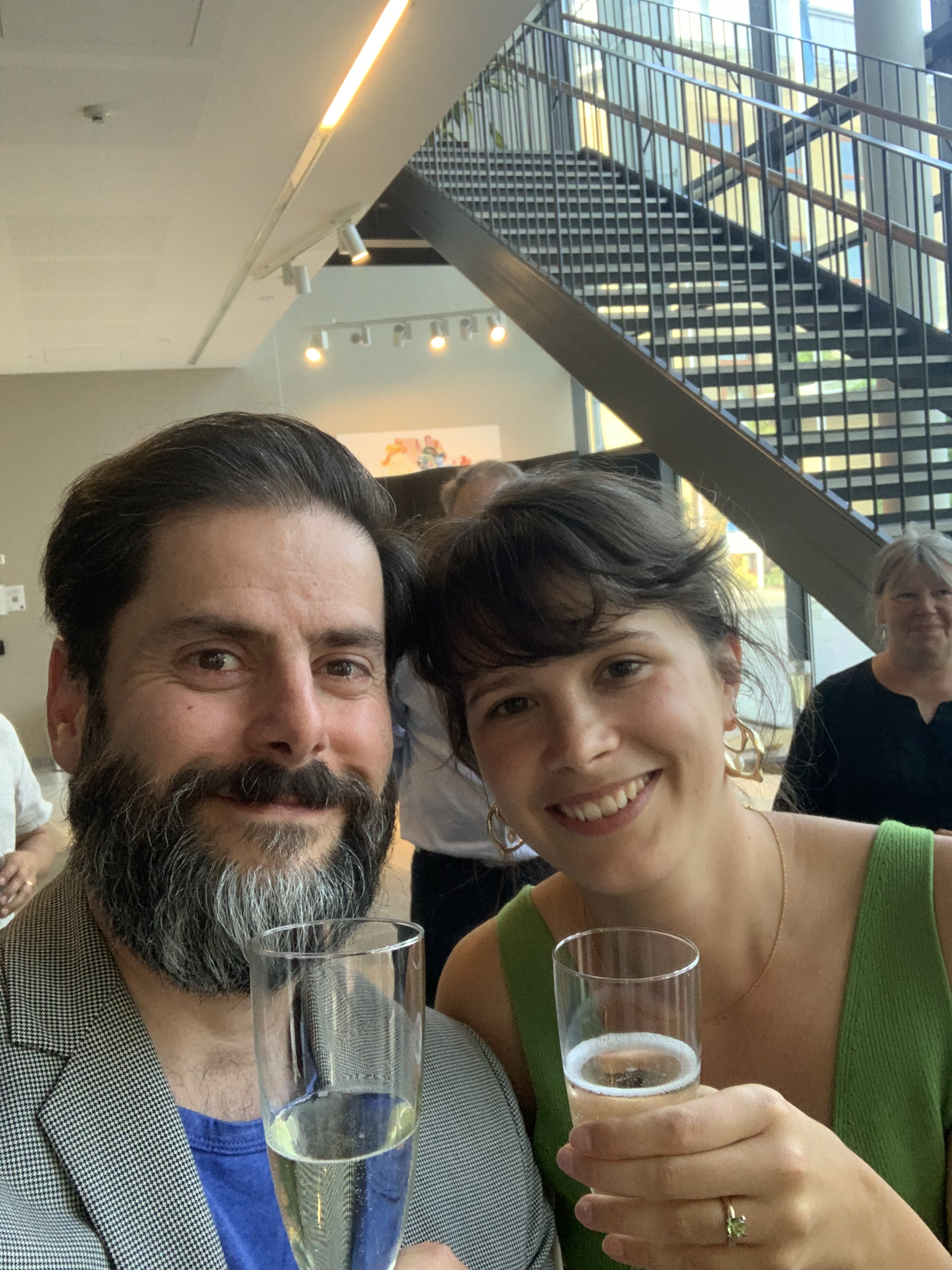
Congratulations, Dr. Jäverfelt!

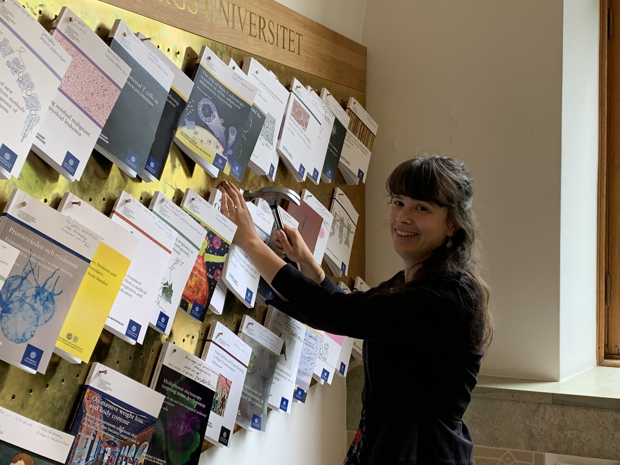
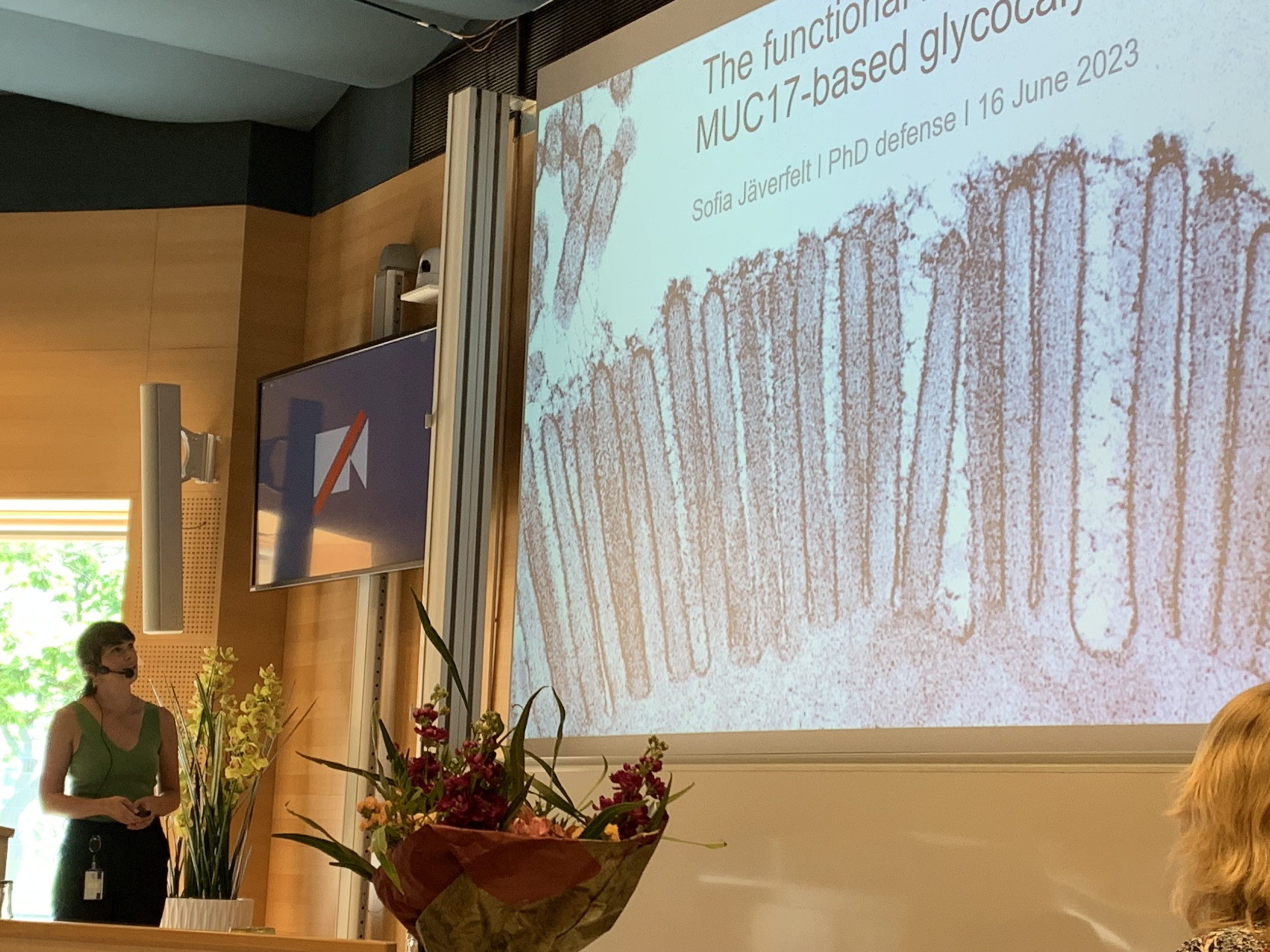
Last Friday, our talented PhD student Sofia successfully and gracefullly defended her thesis on “The functional role of the MUC17-based glycocalyx”!

Sofia’s opponent was Associate Professor Karin Strijbis from Utrecht University.

Congratulations, Dr. Sofia Jäverfelt!

See our work at Universeum Science Center in Gothenburg

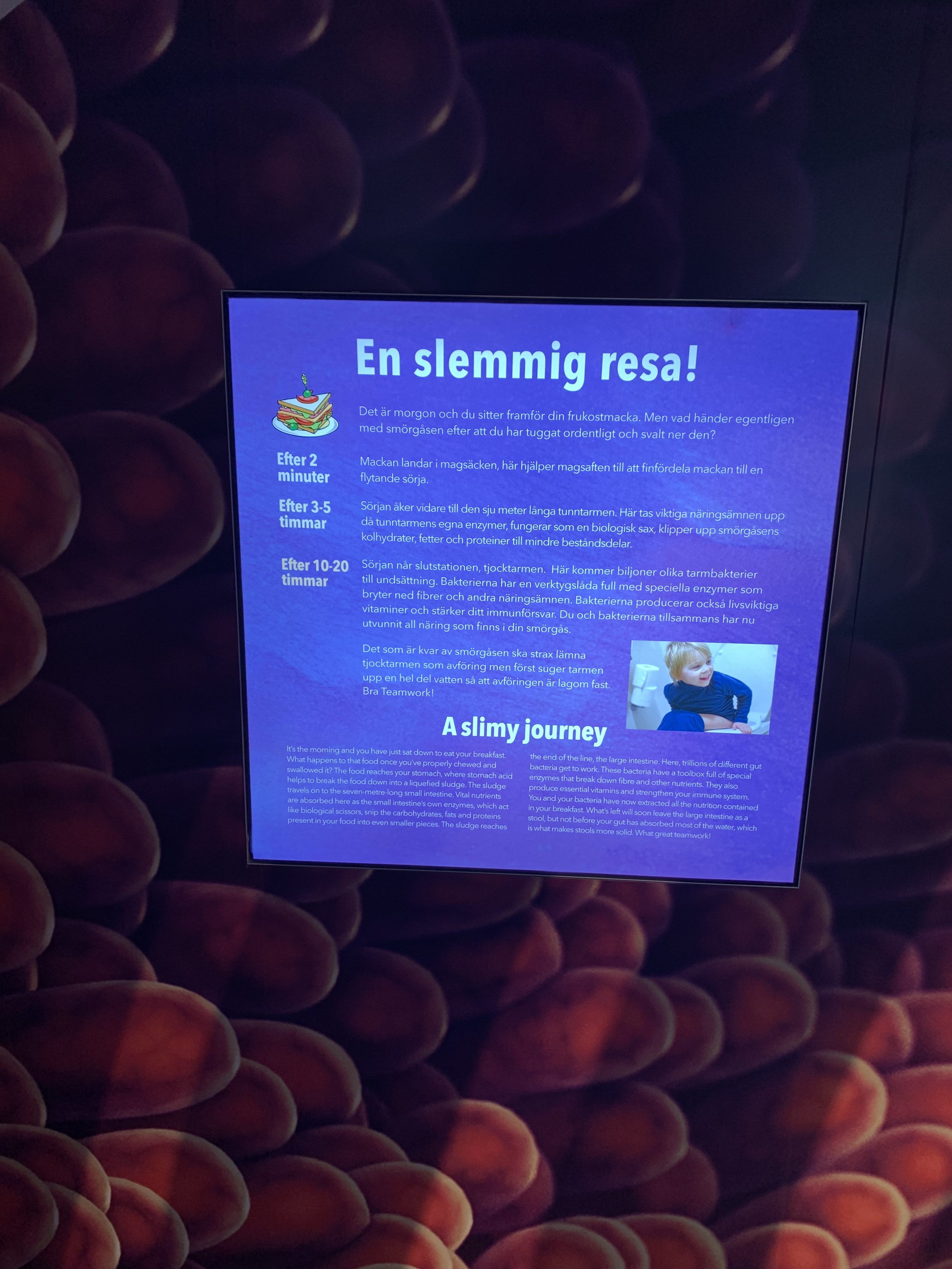
The interactive exhibition Humans opened last week at the science center Universeum in Gothenburg.

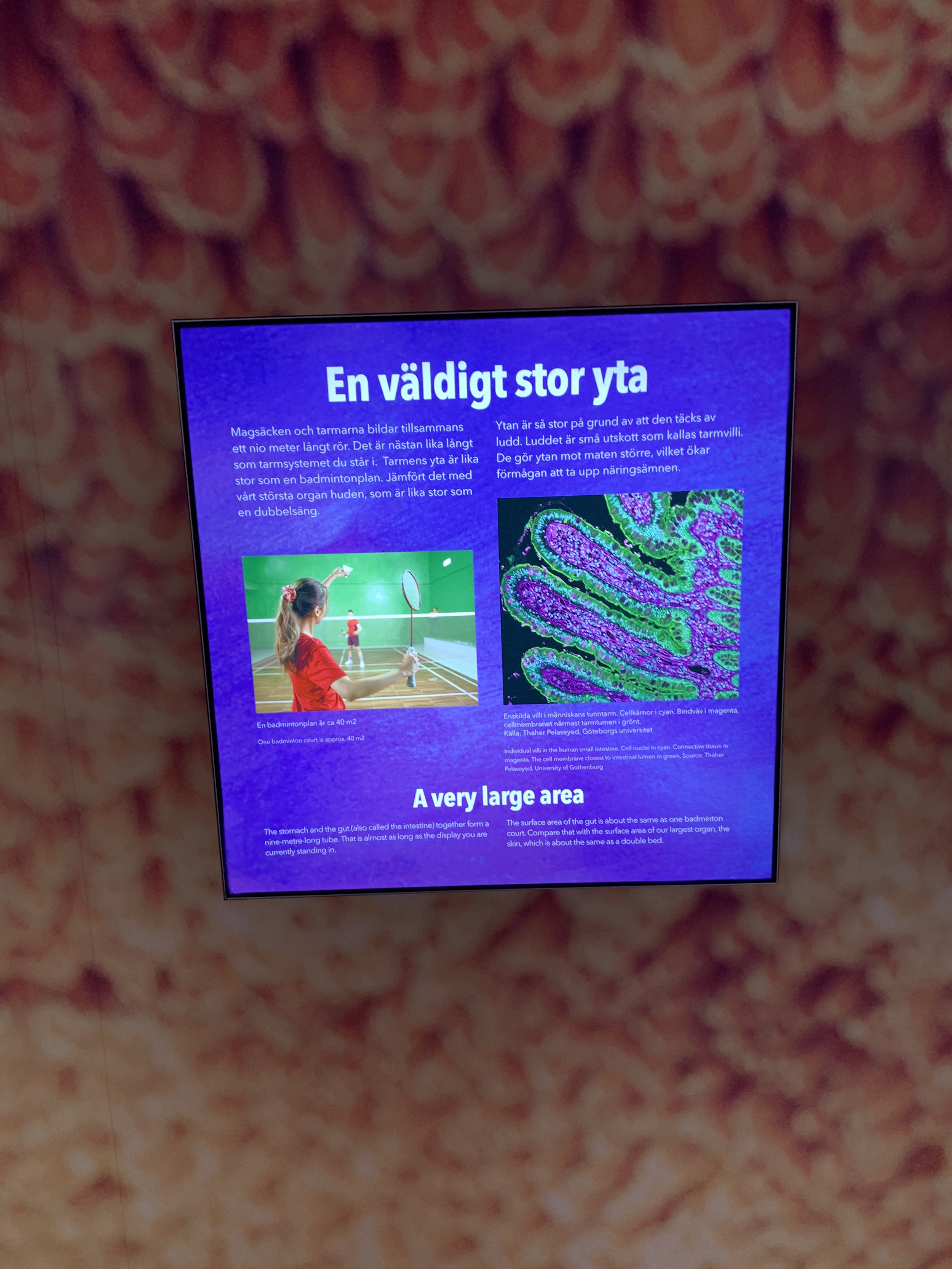
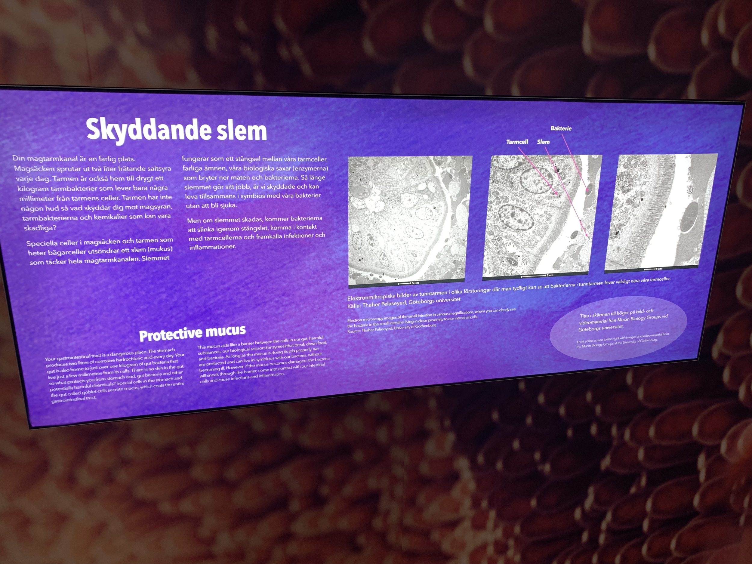
Our lab and other colleagues in the Mucin Biology Groups have curated images, movies and reading material for the exhibition that takes you on a journey through the gastrointestinal tract, where you encounter the fascinating world of mucins, mucus and gut bacteria!

More info about Humans and opening hours here.

Meet Thaher at the Dutch KNVM/NVMM Spring Meeting April 4-5!

Thaher will speak at the Scientific Spring Meeting of the Dutch Society of Medical Microbiology (NVMM) and the Royal Dutch Society of Microbiology (KNVM), held on April 4 and 5, 2023 in Congrescentre Papendal, Arnhem.

Thaher’s talk entitled “From infancy to adulthood: Exploring the barrier function of membrane mucins in the small intestine” is scheduled on Tuesday April 4th, 2 pm.

Read the full program here.

Feature: Mucins help us coexist with bacteria

Interested in our research? Read the latest feature about our research on membrane mucins.

”Fundamentally, we want to understand the principles of human coexistence with trillions of bacteria in our intestines. We as humans are deeply dependent on bacteria, the gut microbiota, for our health. Paradoxically, the same bacteria can turn into vicious enemies if they are not kept in check. Our way of controlling the microbiota is to establish various biological and physical barriers between our tissues and the bacteria. My lab studies the function of membrane mucins that cover the whole length of our small and large intestines. The first membrane mucins were described almost 50 years ago, but their function is still an enigma. We have recently shown that membrane mucins play an important role in protecting our intestinal cells against bacteria. Now, we are starting to realize that they play a much broader role in our coexistence with bacteria. For example, membrane mucins are also present in breast milk, where they present thousands of sugar molecules to the gut microbiota of the infant. These sugar molecules could help select beneficial bacteria or capture and eliminate harmful microbes.”

Read the full feature here.

New paper on discovery of a MUC3B gene in the human genome

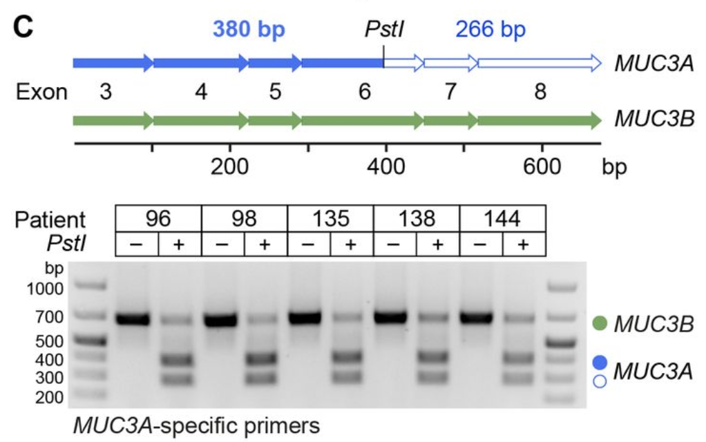
Human tissue surfaces are coated with mucins, a family of macromolecular sugar-laden proteins serving diverse functions from lubrication to the formation of selective biochemical barriers against harmful microorganisms and molecules. Mucin proteins carry long stretches of tandemly repeated sequences that undergo extensive O-linked glycosylation to form linear mucin domains. The repetitive nature of mucin domains makes them prone to recombination and renders their genetic sequences particularly difficult to read with standard sequencing technologies. Our recent work, published in PLOS ONE, leveraged a recent human genome assembly to characterize a previously unmapped MUC3B gene located at the q22 locus on chromosome 7, within a cluster of four structurally related membrane mucin genes that we name the MUC3 cluster.

This work was a collaboration with Tiange Lang at Big Data Decision Institution, Jinan University, China.

Read our paper here.

Compete genomic, mRNA and protein sequences for mucin genes MUC3A, MUC3B, MUC12 and MUC17 in the human MUC3 cluster can be downloaded from the Mucin database (v 2.2).

Elena awarded the "Bollan" Stipend for research on functional role of MUC17!

Elena was awarded a Bollan stipend. From right: Anandi, Elena, Thaher, and Sofia.

Elena has been awarded the Elisabet "Bollan" Lindén Stipend from the Core facilities at Sahlgrenska Academy. Congratulations, Elena!

Project details:

“Inflammatory bowel disease (IBD) is caused by bacteria that reach intestinal epithelial cells (IECs) and thereby trigger inflammation. We recently discovered that membrane mucin MUC17 prevents bacterial attachment to IECs. However, the role of MUC17 in IBD and in mucosal defenses remains unexplored. We have now developed a new Muc17 knockout mouse that helps us understand how IECs combat harmful bacteria and whether defects in MUC17 contribute to IBD etiology. Ultimately, our research could result in new therapeutic approaches to treat IBD.”

The stipend will be used to analyze the barrier function of Muc17 using services offered by Bioinformatics and Data Centre (BDC), Centre for Cellular Imaging (CCI), and Experimental Biomedicine (EBM).

Read the full story here.

Scholarships for researchers from Ukraine

Pelaseyed Lab is dedicated to supporting students, PhD students and postdocs from Ukraine who seek refuge in Sweden following the Russian invasion.

If you are looking for a research lab where you can continue your research in life sciences, don’t hesistate to contact us. We will do our best to provide you with scholarships, lab space and supervision.

You can read more about our ongoing research projects here.


Contact Thaher Pelaseyed thaher.pelaseye[at]medkem.gu.se for more information.

Read our preprint: Discovery of a MUC3B gene reconstructs the membrane mucin gene cluster on human chromosome 7

Human tissue surfaces are coated with mucins, a family of macromolecular sugar-laden proteins serving diverse functions from lubrication to formation of selective biochemical barriers against harmful microorganisms and molecules. Membrane mucins are a distinct group of mucins that are attached to epithelial cell surfaces where they create a dense glycocalyx facing the extracellular environment.

However, the repetitive nature of mucin domains makes them prone to recombination and render their genetic sequences particularly difficult to read with standard sequencing technologies. As a result, human mucin genes suffer from significant sequence gaps that have hampered investigation of gene function in health and disease. Here we leveraged a recent human genome assembly to identify a previously unmapped MUC3B gene located within a cluster of four structurally related membrane mucin genes that we entitle the MUC3 cluster at q22 locus in chromosome 7.

Read our preprint on bioRxiv.

New paper out in Cell Reports!

We are happy to share our new paper in Cell Reports, where we introduce membrane mucin MUC17 as new component of the mutilfaceted intestinal barrier. Elena has led this project, which revealed that MUC17 is associated with an innate immunity repertoire is triggered in the small intestine during the weaning transition. At this crucial event in mammalian development, MUC17 expression is induced in enterocytes, upon which MUC17 localizes to the brush border membrane to function as a barrier that prevents bacteria from binding to the host epithelium.

Congratulations, Elena, and thank you to all lab members and colleagues in the Mucin Biology Groups at University of Gothenburg!

Read our latest review on JCS!

JCS240929supp.jpg

Thaher and Professor Gunnar C. Hansson share their latest thoughts on the function, regulation and the future of membrane mucins of the intestine. Read the abstract below.

“Membrane mucins cover most mucosal surfaces throughout the human body. The intestine harbors complex population of microorganisms (the microbiota) and numerous exogenous molecules that can harm the epithelium. In the colon, where the microbial burden is high, a mucus barrier forms the first line of defense by keeping bacteria away from the epithelial cells. In the small intestine where the mucus layer is less organized, microbes are kept at bay by peristalsis and antimicrobial peptides. Additionally, a dense glycocalyx consisting of extended and heavily glycosylated membrane mucins covers the surface of enterocytes. Whereas many aspects of mucosal barriers are being discovered, the function of membrane mucins remains a largely overlooked topic, mainly because we lack the necessary reagents and experimental animal models to investigate these large glycoproteins. In this Cell Science at a Glance article and accompanying poster, we highlight central concepts of membrane mucin biology and the role of membrane mucins as integral components of intestinal mucosal barriers. We also present the current consensus concerning the role of membrane mucins in host–microbe interactions. Moreover, we discuss how regulatory circuits that govern membrane mucins in the healthy gut display strong overlap with pathways that are perturbed during chronic inflammation. Finally, we review how dysregulation of intestinal membrane mucins may contribute to human diseases, such as inflammation and cancer.”

Read more here.

Our NIH/MIST grant got extended!

Last year our lab received a prestigous 1-year grant from the Mucosal Immunology Studies Team, a NIH-funded consortium. We have just received news that MIST/NIH will extend our funding with an additional year. Now we will continue our work on identifying the external environmental factors that drive membrane mucin expression in the small intestine.

Travel grants to Anandi, Elena and Sofia!

Congratulations to Anandi, Elena and Sofia who all received travel grants for participation in international conferences during 2019!

Anandi and Sofia will attend the 15th International Workshop on Carcinoma-associated Mucins in Cambridge, UK with poster presentations of their work.

Elena will attend the UEG Week (Oct 19-23) in Barcelona, Spain.